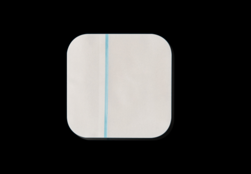
Hydrocolloid wound dressings represent a pivotal development in modern wound care, offering a unique balance of moisture retention, protection, and bioactive wound management. These semi-occlusive or occlusive dressings are especially valued for their ability to maintain a moist wound environment, which is critical for promoting autolytic debridement and accelerating tissue regeneration. Since their introduction in the 1970s, hydrocolloid dressings have been widely adopted in both clinical and home care settings for managing a broad range of acute and chronic wounds.
This article provides an in-depth exploration of hydrocolloid dressing technology, examining their chemical composition, mechanism of action, performance in clinical practice, material innovation, and their role in the broader context of advanced wound management.
1. Composition and Structure of Hydrocolloid Dressings
Hydrocolloid dressings are multilayered composite materials typically composed of:
-
Inner Hydrocolloid Layer: This is a gel-forming matrix usually composed of hydrophilic polymers such as gelatin, pectin, and sodium carboxymethylcellulose (NaCMC). These materials are selected for their ability to absorb exudate and transform into a gel.
-
Outer Backing Layer: Often made of polyurethane film or foam, this layer is waterproof and impermeable to bacteria but may be semi-permeable to water vapor and gases like oxygen and carbon dioxide, helping to manage moisture and reduce maceration risk.
-
Adhesive Components: A pressure-sensitive adhesive binds the dressing to intact skin. It may be integrated into the hydrocolloid matrix or layered separately.
Some hydrocolloid dressings may also contain antimicrobial agents (e.g., silver or honey), foam layers for cushioning, or film overlays to improve conformability and mechanical strength.
2. Mechanism of Action in Wound Healing
The efficacy of hydrocolloid dressings is rooted in their ability to create and maintain an ideal healing environment. The primary mechanisms include:
-
Moisture Retention: Hydrocolloids swell upon absorbing exudate, forming a cohesive gel that covers the wound bed. This moist microenvironment promotes cell migration, angiogenesis, and epithelialization.
-
Autolytic Debridement: The moist conditions support endogenous enzymes that digest necrotic tissue without harming healthy tissue, reducing the need for mechanical or surgical debridement.
-
Barrier Protection: The occlusive backing layer prevents bacterial contamination, reducing infection risk. It also serves as a physical barrier against friction and shear.
-
Thermal Insulation: The semi-occlusive nature helps maintain optimal wound temperature (~37°C), which supports enzymatic activity and cell proliferation.
-
Pain Reduction: The gentle adhesion and cushioning properties reduce trauma during dressing changes and provide relief from mechanical irritation.
These effects collectively contribute to faster healing times and improved patient comfort, particularly in wounds that benefit from moist healing dynamics.
3. Indications and Clinical Applications
Hydrocolloid dressings are suitable for a wide spectrum of wound types, especially those with mild to moderate exudate. Common indications include:
-
Pressure Ulcers (Stage I–II, some Stage III): Effective in preventing and managing early-stage pressure injuries.
-
Leg Ulcers (Venous or Arterial): Maintains moisture without macerating periwound skin.
-
Diabetic Foot Ulcers: May be used with caution under close monitoring due to infection risk.
-
Postoperative Incisions: Helps manage exudate and protect surgical wounds.
-
Superficial Burns and Abrasions: Promotes rapid reepithelialization with minimal discomfort.
-
Donor Sites and Skin Grafts: Provides moisture balance and mechanical protection.
Contraindications include infected wounds, wounds with heavy exudate, deep cavity wounds (unless used with fillers), and wounds requiring frequent inspection.
4. Comparative Performance and Evidence-Based Outcomes
Numerous clinical studies have evaluated hydrocolloid dressings against other modern dressings such as foams, alginates, and hydrofibers. Key findings include:
-
Healing Time: Hydrocolloids often show comparable or faster healing rates for superficial and moderate wounds compared to traditional gauze or semi-permeable films.
-
Pain and Comfort: Patients consistently report lower pain scores due to non-adherent gel formation and fewer dressing changes.
-
Cost-Effectiveness: Although more expensive per unit than gauze, the extended wear time (up to 5–7 days) and reduced dressing changes can lower total treatment costs.
-
Infection Control: Standard hydrocolloids are not inherently antimicrobial, but silver-impregnated versions show improved outcomes in mildly colonized wounds.
A Cochrane review of hydrocolloid dressings in pressure ulcer treatment found that while no single dressing was significantly superior in healing rates, hydrocolloids offered clear advantages in patient acceptability and dressing handling.
5. Materials Science and Advances in Hydrocolloid Formulations
Recent years have seen advances in the material design of hydrocolloid dressings, driven by the need for enhanced functionality and biocompatibility.
-
Nanocomposite Hydrocolloids: Incorporating nanomaterials like silver nanoparticles or zinc oxide enhances antimicrobial action without compromising biocompatibility.
-
Bioactive Additives: Some formulations include growth factors, collagen, or honey to accelerate the healing process.
-
Smart Hydrocolloids: Emerging designs integrate pH indicators or colorimetric sensors that change color in response to infection or excessive exudate, allowing for early intervention.
-
Layered Architecture: Multi-layered designs offer compartmentalized functionality—e.g., a gel-forming layer for moisture control, a foam core for absorption, and a film layer for barrier protection.
-
Sustainable and Biodegradable Materials: Driven by environmental concerns, research is exploring the use of bio-based hydrocolloids derived from alginate, chitosan, or cellulose.

6. Role in Modern Wound Care Protocols
Hydrocolloid dressings are integral to modern wound care algorithms, especially under frameworks such as the TIME principle:
-
Tissue Management: Hydrocolloids support autolytic debridement and tissue granulation.
-
Infection and Inflammation Control: Though not inherently antimicrobial, they form a barrier that limits contamination.
-
Moisture Balance: Their gel-forming properties optimize exudate management.
-
Edge of Wound Advancement: By maintaining a moist and insulated environment, hydrocolloids encourage epithelial cell migration.
In practice, hydrocolloids are often used in rotation with other dressing types depending on wound progression. For example, alginate or foam dressings may be used in the early exudative phase, with hydrocolloids applied during the proliferative and maturation phases.
7. User Considerations and Clinical Challenges
Despite their many advantages, hydrocolloid dressings present certain limitations and risks:
-
Occlusion and Anaerobic Infection Risk: The occlusive nature may exacerbate anaerobic bacterial growth in infected wounds.
-
Skin Maceration: Prolonged exposure to gel in poorly managed wounds can lead to maceration of surrounding skin.
-
Allergic Reactions: Some patients may develop contact dermatitis or adhesive sensitivity.
-
Transparency and Monitoring: Hydrocolloids are generally opaque, making wound inspection difficult without removing the dressing.
-
Removal Challenges: Adhesion to dry or fragile skin can cause trauma; removal techniques must be gentle and systematic.
Proper wound assessment, regular monitoring, and practitioner training are critical to ensuring optimal outcomes with hydrocolloid therapy.
8. Regulatory and Safety Considerations
Hydrocolloid dressings are classified as medical devices and are regulated under frameworks such as:
-
FDA 510(k) Clearance (USA): Requires demonstration of substantial equivalence to a legally marketed device.
-
CE Marking (EU): Ensures compliance with MDR (Medical Device Regulation) for safety and performance.
-
ISO 10993: Governs biocompatibility testing including cytotoxicity, irritation, and sensitization.
Packaging and labeling must include indications, contraindications, shelf life, storage conditions, and disposal instructions. Sterile versions must meet ISO 11137 standards for sterilization (e.g., gamma radiation or ethylene oxide).
9. Future Directions and Research Opportunities
The future of hydrocolloid wound dressing technology lies at the intersection of material science, biotechnology, and digital health. Key areas of ongoing research include:
-
Antimicrobial and Anti-Biofilm Agents: Development of formulations that can actively disrupt biofilms or resist colonization.
-
Smart Wound Monitoring: Integration of biosensors for pH, moisture, temperature, and enzymatic activity within the dressing matrix.
-
3D Printing of Custom Dressings: Personalized hydrocolloid designs tailored to wound morphology and exudate profile.
-
Hybrid Systems: Combinations with other dressings (e.g., foam-hydrocolloid, alginate-hydrocolloid) to merge functionalities.
-
Machine Learning for Dressing Selection: Data-driven systems that predict optimal dressing choices based on patient profiles and wound characteristics.
-
Eco-Friendly Solutions: Use of renewable materials and recyclable packaging to reduce environmental impact.
Clinical translation of these technologies depends on cost-effectiveness, regulatory approval, manufacturing scalability, and healthcare provider education.

 English
 中文简体
中文简体


