
 English
 中文简体
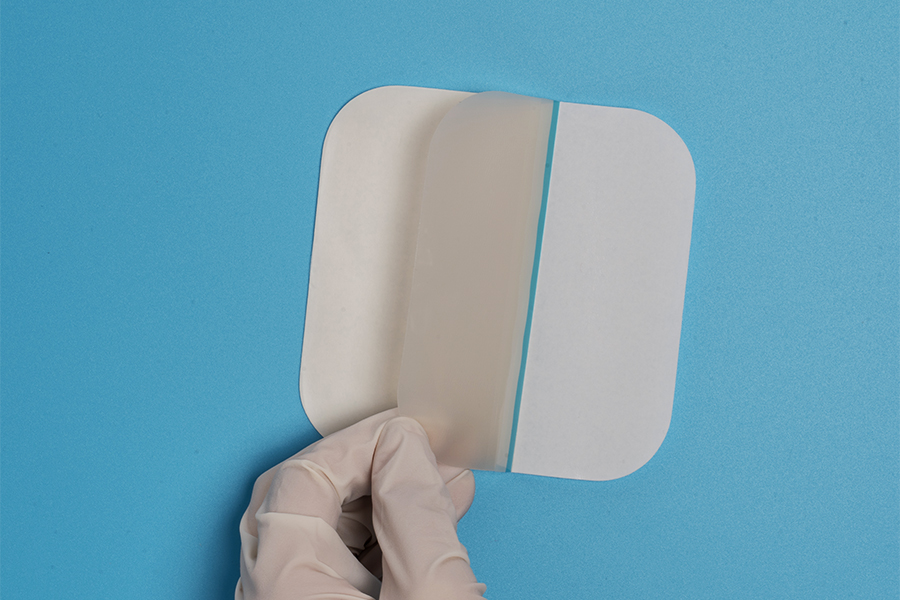
中文简体In the realm of modern wound care, hydrocolloid dressings stand as a paradox: unassuming in appearance yet revolutionary in function. These gel-forming patches, often resembling translucent stickers, have transformed the treatment of chronic and acute wounds by leveraging the power of moisture—a concept that defied traditional "dry healing" dogma for centuries. How do these dressings accelerate tissue regeneration while preventing infection? What hidden complexities lie within their sticky matrices? This 4,000-word exploration dissects hydrocolloid technology, from its serendipitous origins in food science to its role in cutting-edge regenerative medicine, while confronting controversies over cost, sustainability, and clinical efficacy.
1. The Evolution of Hydrocolloid Dressings: From Starch to Smart Healing
Historical Roots in Adhesives and Food Science
The story begins not in a medical lab but in a 19th-century kitchen. Chemists experimenting with starch-based adhesives observed that mixtures of gelatin, pectin, and water formed flexible, moisture-retaining films. By the 1930s, these hydrocolloids (water-loving colloids) were used to seal canned goods. The leap to medicine came in 1962 when Dr. George Winter’s landmark study on moist wound healing overturned centuries of belief that wounds healed best when dried.
Key Milestones
-
1970s: ConvaTec introduces DuoDERM®, the first commercial hydrocolloid dressing, combining sodium carboxymethylcellulose (CMC) and elastomers.
-
1980s: Incorporation of polyurethane films enhances breathability and conformability.
-
2000s: Antimicrobial additives (silver, honey) address biofilm resistance.
-
2020s: "Smart" hydrocolloids with pH sensors and drug-eluting capabilities emerge.
Cultural Shifts
Hydrocolloids democratized advanced wound care, enabling home management of ulcers and burns previously requiring hospitalization. Their adoption also spurred debates on medicalization—turning minor wounds into "conditions" needing specialized products.
2. Material Science: Decoding the Hydrocolloid Matrix
Core Components
A hydrocolloid dressing is a carefully engineered ecosystem:
-
Absorbent Particles: Sodium CMC, gelatin, pectin, or alginate. These hydrophilic polymers swell into a gel upon fluid absorption.
-
Elastomeric Backing: Polyurethane or polyisobutylene provides flexibility and occlusivity.
-
Adhesive Layer: Skin-friendly acrylates or silicone ensure adhesion without trauma.
-
Functional Additives:
-
Glycerin: Plasticizes the gel, preventing crust formation.
-
Silver ions: Combat Pseudomonas and MRSA biofilms.
-
Collagen: Enhances fibroblast migration.
-
The Gelation Process
When wound exudate contacts the dressing:
-
Hydration: Sodium CMC fibers unwind, absorbing 10–15 times their weight in fluid.
-
Gel Formation: A viscous, cohesive gel forms, maintaining a pH of 4.5–6.5 (optimal for healing).
-
Autolytic Debridement: Moisture softens necrotic tissue, which is enzymatically broken down.
Why Hydrocolloids Outperform Gauze
-
Moisture Balance: Prevents maceration (excess fluid) and desiccation (drying).
-
Thermal Insulation: Maintains a 37°C microenvironment, accelerating cell proliferation.
-
Barrier Function: Occlusive layer blocks bacteria but allows O₂/CO₂ exchange (≈500 g/m²/day MVTR).
3. Clinical Applications: Beyond Band-Aids
A. Chronic Wounds
-
Diabetic Foot Ulcers (DFUs): Hydrocolloids reduce healing time by 40% compared to saline gauze (RCT data: Diabetes Care, 2018).
-
Pressure Injuries: Gel cushions redistribute pressure while absorbing exudate.
B. Acute Wounds
-
Partial-Thickness Burns: A Cochrane review (2021) found hydrocolloids reduce pain during dressing changes by 60%.
-
Surgical Incisions: Post-cesarean studies show lower infection rates vs. traditional sutures.
C. Dermatology
-
Blister Management: Fluid absorption prevents rupture in friction blisters.
-
Psoriasis/ Eczema: Hydrocolloids with ceramides restore skin barrier function.
D. Pediatric and Geriatric Care
-
Gentle removal minimizes trauma to fragile skin.
-
Transparent variants allow monitoring without disturbing the wound.
4. The Manufacturing Alchemy: From Lab to Patient
Production Process
-
Mixing: Hydrocolloid powders (CMC, gelatin) are dissolved in purified water under vacuum to avoid air bubbles.
-
Coating: The slurry is spread onto silicone-coated release liners at 100–200 µm thickness.
-
Lamination: Elastomeric backing films are heat-pressed onto the gel layer.
-
Die-Cutting: Dressings are cut into shapes (oval, square, island) using laser or rotary dies.
-
Sterilization: Gamma irradiation (25–40 kGy) ensures microbial safety without degrading polymers.
Quality Control Challenges
-
Gel Integrity Testing: Ensures the dressing withstands 300% elongation without tearing.
-
Fluid Handling Capacity: Measured via ASTM F2818-10 (grams of saline absorbed per cm²).
-
Adhesion Balance: Peel adhesion force (measured in N/25mm) must be strong enough to seal but weak enough to avoid skin stripping.
Cost Drivers
-
Raw Materials: Medical-grade CMC costs 5/kg for industrial grade.
-
Regulatory Compliance: FDA 510(k) clearance adds 2M per product.
5. Controversies and Limitations
A. Clinical Debates
-
Infection Risks: Occlusive environments may favor anaerobic bacteria (Clostridium spp.) in heavily exuding wounds.
-
Allergic Reactions: 12% of patients develop contact dermatitis to acrylate adhesives (study: Contact Dermatitis, 2020).
-
Overuse: Inappropriate application to dry or necrotic wounds delays healing.
B. Environmental Impact
-
Non-Recyclable Waste: Used dressings, contaminated with biohazards, contribute to 2.5 million tons of medical waste annually.
-
Microplastic Shedding: Polyurethane backings degrade into <5mm particles, infiltrating waterways.
C. Economic Barriers
A hydrocolloid dressing costs 0.50 for gauze—a critical issue in low-income countries where diabetic ulcers prevalence is highest.
6. Innovations Redefining Hydrocolloid Technology
A. Bioactive Upgrades
-
Stem Cell Attractants: Dressings infused with SDF-1α chemokine recruit endogenous stem cells.
-
Electrospun Fibers: Nano-fibrous hydrocolloids mimic extracellular matrix structure.
-
Quorum Sensing Inhibitors: Disrupt bacterial communication without antibiotics.
B. Sustainability Solutions
-
Biodegradable Backings: Poly(lactic-co-glycolic acid) (PLGA) films decompose in 6–12 months.
-
Plant-Based Hydrocolloids: Cactus mucilage and okra polysaccharides replace synthetic polymers.
-
Closed-Loop Recycling: Autoclaving recovers silver ions for reuse.
C. Digital Integration
-
Smart Sensors: RFID tags monitor exudate pH and temperature, alerting clinicians to infection via Bluetooth.
-
3D Printing: Patient-specific dressings conform to complex anatomies (e.g., facial burns).
7. Global Market Dynamics and Future Projections
Market Growth
Valued at 3.2 billion by 2030 (CAGR 8.5%), driven by:
-
Aging populations (23% of Europe will be >65 by 2030).
-
Diabetes pandemic (700 million cases expected by 2045).
Regional Disparities
-
North America/Europe: Dominated by premium antimicrobial products.
-
Asia-Pacific: Fastest growth due to medical tourism and rising healthcare expenditure.
-
Africa: Limited access; 80% of wounds still treated with traditional methods.
The Road Ahead
-
Personalized Dressings: Genomic profiling to match dressing composition with patient’s inflammatory markers.
-
Space Medicine: NASA-funded research on hydrocolloids for zero-gravity wound management.
-
Ethical AI: Algorithms to optimize dressing selection, reducing trial-and-error in clinics.


")


